We offer radiological services at New London Hospital (NLH) and the Newport Health Center. We work with providers at Dartmouth Hitchcock Medical Center as well.
Our technologists are registered, certified radiologic technologists. Our board-certified radiologists interpret all images and tests. We offer routine radiology services, such as:
- 3-dimensional (3D) tomosynthesis digital mammography
- Bone mineral density testing
- Computed tomography (CT) scans
- Magnetic resonance imaging (MRI)
- Nuclear medicine
- Ultrasound
3D tomosynthesis digital mammography
3D mammography offers greater flexibility and magnification for women with denser breast tissue. Breast images produced through computerization rather than X-ray film are clear, easy to read, and are ready to view within seconds. NLH also uses a computer-aided detection system. This provides a computerized analysis of a patient’s mammogram in addition to the radiologist’s interpretation. The 3D tomosynthesis technology improves the detection of cancer. It may help reduce the need for repeat mammograms. Our technologists are certified in mammography.
Bone mineral density testing
A bone density test called DEXA scan is a test that can diagnose osteoporosis before a broken bone occurs. This test helps estimate the density of your bones and your chance of breaking a bone. The procedure is painless and safe and usually takes 10 to 15 minutes. To prepare for your DEXA Scan, dress comfortably wearing clothes without metal buttons, buckles, or zippers. We provide bone mineral density scans every Monday through Friday.
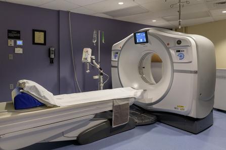
CT scans
CT scans use X-rays to make detailed pictures of structures inside of the body.
During the test, patients lie on a table attached to the CT scanner. The CT scanner is a large doughnut-shaped machine. The CT scanner sends X-rays through the selected body area. Each rotation of the scanner takes less than a second and provides a picture of a thin slice of the organ or area.
An iodine dye (contrast material) is often used to make structures and organs easier to see on CT images. We use the dye to check blood flow, find tumors, and look for other problems. The dye may be put in a vein in the arm, using an IV or it may be placed into other parts of the body (such as the rectum or a joint) to see those areas better. For some types of CT scans patients are asked to drink the dye. CT pictures are taken before and after the dye is used.
Our CT scanner is available 24 hours a day, 7 days a week. The scanner is available for a wide range of imaging. Our technologists are all certified in CT.
MRI
An MRI is a test that uses a magnetic field and pulses of radio wave energy to make pictures of organs and structures inside the body. Our technologists are all certified in MRI. For an MRI test, our technologists place the selected area of the body inside a special machine that contains a strong magnet. Often, MRI scans give different information about structures in the body than with an X-ray, ultrasound, or CT scan. MRI scans also may show problems that other imaging methods may not show.
Nuclear medicine
Nuclear medicine is a branch of radiology that uses radioactive materials to determine if certain organs are working properly. Nuclear medicine is used to examine various organs, such as:
- Bones (for cancer, infection, or trauma)
- Brain
- Heart
- Kidneys
- Liver
- Lungs
- Thyroid
Before the test begins, patients receive a small amount of radioactive material called a radioisotope. This radioisotope may be swallowed or given by injection. In many cases, a delay exists between the time the material is administered and the test begins. This allows the isotope to flow through the body and concentrate in the organ that is being examined.
Our providers track the isotope as it moves through the organ. This provides valuable information about how the selected organ is functioning. In most studies, the patient lies on a comfortable table. A large camera is used to scan the body. The camera moves or rotates around the patient.
Nuclear medicine services are available every Monday. Our technologists are certified in nuclear medicine.
Ultrasound
Ultrasound images help our providers diagnose a wide range of diseases and conditions. An ultrasound test transmits high-frequency sound waves through body tissues. Using an instrument called a transducer, the ultrasound machine transfers the information to a computer. The provider can then see the ultrasound images on a monitor to help in diagnosis.
Radiology team