


We offer radiological services at New London Hospital (NLH) and the Newport Health Center. We work with providers at Dartmouth Hitchcock Medical Center as well.
Our technologists are registered, certified radiologic technologists. Our board-certified radiologists interpret all images and tests. We offer routine radiology services, such as:
- 3-dimensional (3D) tomosynthesis digital mammography
- Bone mineral density testing
- Computed tomography (CT) scans
- Magnetic resonance imaging (MRI)
- Nuclear medicine
- Ultrasound
3D tomosynthesis digital mammography
3D mammography offers greater flexibility and magnification for women with denser breast tissue. Breast images produced through computerization rather than X-ray film are clear, easy to read, and are ready to view within seconds. NLH also uses a computer-aided detection system. This provides a computerized analysis of a patient’s mammogram in addition to the radiologist’s interpretation. The 3D tomosynthesis technology improves the detection of cancer. It may help reduce the need for repeat mammograms. Our technologists are certified in mammography.
Bone mineral density testing
A bone density test called DEXA scan is a test that can diagnose osteoporosis before a broken bone occurs. This test helps estimate the density of your bones and your chance of breaking a bone. The procedure is painless and safe and usually takes 10 to 15 minutes. To prepare for your DEXA Scan, dress comfortably wearing clothes without metal buttons, buckles, or zippers. We provide bone mineral density scans every Monday through Friday.
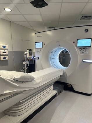
CT scans
CT scans use X-rays to make detailed pictures of structures inside of the body.
During the test, patients lie on a table attached to the CT scanner. The CT scanner is a large doughnut-shaped machine. The CT scanner sends X-rays through the selected body area. Each rotation of the scanner takes less than a second and provides a picture of a thin slice of the organ or area.
An iodine dye (contrast material) is often used to make structures and organs easier to see on CT images. We use the dye to check blood flow, find tumors, and look for other problems. The dye may be put in a vein in the arm, using an IV or it may be placed into other parts of the body (such as the rectum or a joint) to see those areas better. For some types of CT scans patients are asked to drink the dye. CT pictures are taken before and after the dye is used.
Our CT scanner is available 24 hours a day, 7 days a week. The scanner is available for a wide range of imaging. Our technologists are all certified in CT.
CT Lung Screenings
NLH Radiology meets the high standards in quality and safety to be a designated Lung cancer screening center. CT Lung cancer screening is a noninvasive, low-dose scan to find lung cancer in its early stages. Early detection increases the chance of successful treatment. If you are age: 50 to 80 years old and are a current or former smoker with no signs or symptoms, check with your health care provider to see if you are eligible for these screenings.
MRI
An MRI is a test that uses a magnetic field and pulses of radio wave energy to make pictures of organs and structures inside the body. Our technologists are all certified in MRI. For an MRI test, our technologists place the selected area of the body inside a special machine that contains a strong magnet. Often, MRI scans give different information about structures in the body than with an X-ray, ultrasound, or CT scan. MRI scans also may show problems that other imaging methods may not show.
Nuclear medicine
Nuclear Stress Tests
Nuclear Stress tests offered at New London Hospital through a mobile company, Catalyst MedTech. This is the most common type of nuclear cardiology test. It involves taking images before and after a stress phase (exercise or medication) to see how blood flows to the heart muscle. Currently available the 1st and 3rd Wednesday of the month (subject to change).
Ultrasound
Ultrasound images help our providers diagnose a wide range of diseases and conditions. An ultrasound test transmits high-frequency sound waves through body tissues. Using an instrument called a transducer, the ultrasound machine transfers the information to a computer. The provider can then see the ultrasound images on a monitor to help in diagnosis.